

New UMBC research reveals how two different parts of the hippocampus—the brain’s memory center—team up in a key reward region to help mice, and likely humans, combine memories of places and contexts with the drive to pursue rewards.

The findings offer fresh insight into how the brain integrates information about “where” and “what feels good” to guide everyday decisions, such as heading to a favorite restaurant to meet friends or seeking out rewarding experiences. Specifically, this discovery, published in the Journal of Neuroscience, shows that inputs from the dorsal and ventral hippocampus converge on the same individual neurons in another brain region, the nucleus accumbens, where they interact in ways that amplify each other’s effects.
“The connection between the hippocampus and nucleus accumbens is where the brain’s map of where to go meets a sense of why it’s worth going,” explains senior author Tara LeGates, assistant professor in UMBC’s Department of Biological Sciences.
For years, scientists viewed the connections from the dorsal hippocampus, which is more closely tied to spatial memory and navigation, and the ventral hippocampus, which is linked more strongly to emotions and motivation, as mostly separate. This paper challenges that understanding.
“A single neuron can receive inputs from different brain regions, and figuring out how it integrates them is crucial for understanding what drives goal-directed actions,” LeGates says.
While the current study focuses on individual cells, the implications reach further. Better knowledge of how these reward-related circuits process and combine information could shed light on conditions where motivation is disrupted, such as depression, addiction, or anxiety disorders.
A close-up on convergence
The research team used advanced methods including using light to stimulate specific pathways (a technique called optogenetics), precise recordings of electrical activity in neurons, and detailed microscope imaging to identify a group of neurons in a specific part of the accumbens that receives direct input from both the dorsal and ventral hippocampus.
Importantly, the synapses involved in these two pathways sit very close together—often within a couple of microns (thousandths of a millimeter)—on the same branches of the neurons’ dendrites, which look like tree roots on nerve cells. That proximity allows them to influence each other quickly. The team found that when both inputs are active at the same time, they produce a stronger combined response than either one alone.

The researchers collaborated with Tagide deCarvalho, director of UMBC’s Keith Porter Imaging Facility, to obtain the high-resolution imaging that confirmed these close partnerships. Upgraded software at the facility allowed the team to capture ultra-thin digital slices (0.2 microns thick) and create 3D reconstructions of neuron branches, clearly demonstrating the close proximity of the synapses that would allow them to interact.
The study’s first author, Ashley Copenhaver ’20, mathematics and biological sciences, Ph.D. ’25, neuroscience and cognitive sciences, led much of the hands-on work in recordings and imaging while mentoring undergraduate team members.

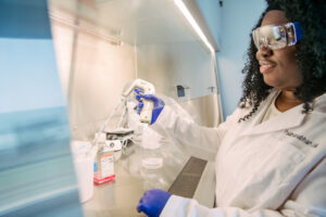
“One of the most exciting parts of this technically challenging project was performing dual-color optogenetics during electrophysiology—I was literally shining tiny beams of red and blue light onto brain tissue, which was activating the dorsal or ventral hippocampus neurons, so that I could record the electrical responses in the nucleus accumbens neurons. It was magical,” Copenhaver says. “Beyond loving the technique, in my opinion, we identified some really critical and fundamental mechanisms of signal integration within the brain. I’m super excited to see where this work heads next.”
From cells to behavior
Understanding how a single neuron handles signals from different brain areas is key to grasping complex behaviors, says LeGates, who has a secondary appointment in the Department of Pharmacology and Physiology at the University of Maryland School of Medicine. Signals from the dorsal and ventral hippocampus are “probably converging more than we’ve previously appreciated, which could change how people approach questions about motivation and learning,” she adds.
That kind of convergence likely helps animals form associations between rewarding outcomes and the environments where they occur—an essential capability for survival. Similar convergence has been seen in other brain areas involved in emotional learning, LeGates says, suggesting the brain may use this strategy widely to link a particular context with feeling and action.

LeGates’ lab is already building on this paper’s foundation by exploring how stress and substances like food, medications, and illicit drugs affect these same connections, with the long-term aim of informing more targeted treatments for various mental health conditions. In the immediate future, the team hopes to record activity from these specially connected neurons during real behaviors to directly link the newly discovered crosstalk between the ventral and dorsal hippocampus to actions.
By uncovering this hidden layer of cooperation between hippocampal pathways, the LeGates lab has advanced our understanding of how the brain weaves together memory and motivation—a fundamental process that shapes the decisions driving daily life.