We use cookies for analytics and, unless you opt out, advertising.
You can opt out of advertising at any time.UMBC uses essential cookies to run this site. With your permission we would also use analytics cookies (to understand how the site is used) and advertising cookies (to measure and target our ads on other platforms). Nothing else loads unless you accept. You can change your choice any time from Cookie Settings in the footer.Privacy Policy
EssentialRequired for the site to work (login, security, your cookie choice). Always on.
UMBC has received a $1.2 million solar energy grant from the Maryland Energy Administration (MEA) to support solar power installations on campus and other sustainability initiatives.
The funding will enable construction of solar canopies over the north portion of the Stadium Lot. Rooftop solar arrays will be mounted on UMBC’s central receiving warehouse, located adjacent to the lot. The installations will be highly visible to the UMBC community and members of the public attending events at the Chesapeake Employers Insurance Arena.
Together, these solar installations will generate 1,000 kWAC of clean, carbon-free energy, or about 2.5 percent of UMBC’s current annual electricity. The solar panels’ output will reduce UMBC’s carbon footprint by roughly 500 tons per year and earn Solar Renewable Energy Credits from the state of Maryland, which can be sold to power companies. The combined savings from the electricity generation and accompanying SRECs may save UMBC $200,000 to $300,000 annually.
“This solar energy project is a significant step forward for UMBC to reach its sustainability goals, and will benefit our campus and local communities,” Taylor Smith, assistant director in the Office of Sustainability, says. “For the first time, the university will generate a significant amount of clean, renewable electricity right here on campus. We are lucky to have a community of partners that made this happen.”
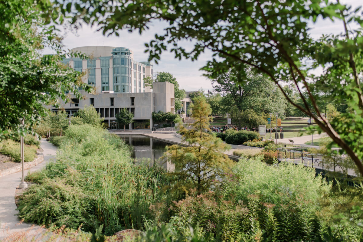
The UMBC Library Pond and surrounding vegetation are home to numerous wildlife species, such as yellow-crowned night herons and red-winged blackbirds. The pond also serves as an important stormwater management feature on campus. (Marlayna Demond ’11/UMBC)
Beyond solar
The grant will also support the development of UMBC’s Campus Clean Energy Master Plan (CCEMP). The CCEMP will rely on past studies and a forthcoming decarbonization engineering study to identify opportunities to save energy and decarbonize campus energy systems, including the central and satellite utility plants.
UMBC’s geography and environmental systems department and engineering faculty affiliated with the Grand ChallengesScholars Program are designing new learning opportunities that will use the solar project as an educational tool. Grant funds will partially fund academic experiences that provide students with hands-on learning opportunities in solar energy technology, renewable energy systems, and sustainability management, integrating real-world problem-solving into the academic curriculum. Five interns will also gain critical experience through supporting these projects, under the supervision of the Office of Sustainability and departmental faculty.
“The campus community can be proud of this commitment to bring solar energy to campus,” Smith says. “This project will not only accelerate UMBC’s shift to clean energy but will also create a visible, tangible symbol of the progress that’s been made. Plus, the new solar arrays will create learning opportunities for students to build the skills they need to thrive in the green economy.”
Thomas Seidman, late emeritus professor of mathematics and statistics, gave 45 years of his life to teaching, mentoring, and conducting research at UMBC before retiring in 2017. After his death in August 2024, his estate donated $1.06 million to UMBC to create the Dr. Thomas I. Seidman Endowed Chair in mathematics. The Maryland E-nnovation Initiative, an effort within the Maryland Department of Commerce, matched the bequest, bringing the total endowment to more than $2 million.
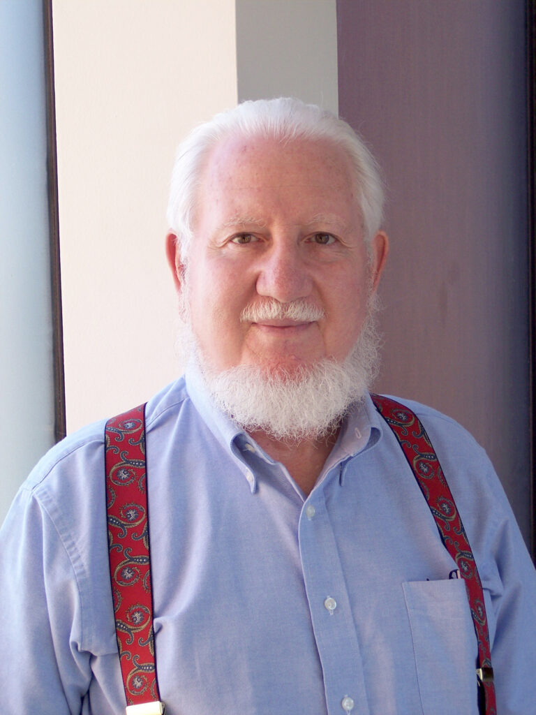
Thomas Seidman (Courtesy Seidman family)
“The Seidman family’s generous gift, along with the MEI match, will make it possible for UMBC to hire world-class applied mathematics faculty with expertise in research fields that will drive the economy of the future,” said William R. LaCourse, dean of the College of Natural and Mathematical Sciences. “CNMS is deeply grateful for Dr. Seidman’s service to UMBC throughout his decades as a faculty member. With this gift, his impact will extend even further by helping UMBC students prepare for rewarding careers in booming fields like data science and AI.”
Joining UMBC only a few years after the university’s founding in 1966, Seidman created a home for himself in the mathematics and statistics department. In addition to his prolific and widely-cited scholarship in applied analysis and fierce dedication to teaching, Seidman also contributed to the young department’s development through writing bylaws for department chair election processes, chairing the promotion and tenure committee for many years, and serving as acting department chair in 1992. He never missed a department seminar and was known for asking insightful questions.
“He was able to build a niche and a role for himself that fit him perfectly,” Seidman’s son, Gregory Seidman, says. “The continuity and stability of the environment, combined with the roots our family had put down in the area, made him feel at home in a way he couldn’t imagine rebuilding elsewhere.”
Leaving a legacy
Seidman “cared deeply about teaching,” his son says, and took pride in instructing students in both foundational math courses like Linear Algebra and courses for non-majors, such as a course on the history of mathematics. Seidman’s one regret was that he did not advise more graduate students, his son shares.
Gregory believes his father hoped to leave a legacy by supporting further work in his mathematical research fields and creating opportunities for students. “It is my hope that his bequest will support many doctoral students following in his research footsteps,” Gregory says, “who would otherwise have been that legacy.”
The younger Seidman remembers spending much of his childhood in his father’s office and in a UMBC computer lab. Father and son learned to program an original Apple PC together in the early 1980s, which inspired Gregory to pursue computer science. Eventually, he even co-authored a research paper with his father. Gregory also recalls regular meetings of the minds with department members in the Seidman household when he was young.
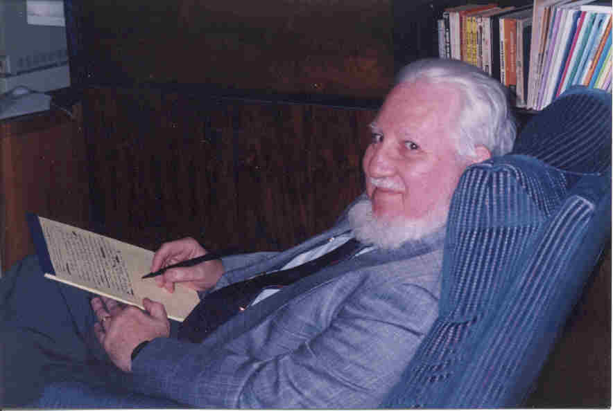
Thomas Seidman was known for his commitment to teaching, and his bequest will create more opportunities for UMBC students to study with outstanding mathematicians. (Courtesy Seidman family)
“While I know my father made a difference at UMBC in various ways, some more significant than others, it feels good to have his name on something that will be seen for years to come,” Gregory says. “I’m also especially pleased that, some day, my kids will be able to show their kids that their great-grandfather, whom they will never meet, made a difference here.”
Seidman notes that his father’s life was a fulfilling one, rich in warm personal relationships and professional success. “His was a life well lived,” he says. And now, the Dr. Thomas I. Seidman Endowed Chair will help create opportunities for more aspiring mathematicians to build their own lives and legacies.
A new $7.8 million award from the Defense Advanced Research Projects Agency (DARPA) will support the development of biologically-powered underwater sensors. Right now, a vast network of underwater sensing devices in oceans around the world conducts environmental monitoring and supports national security—and most of these devices rely on batteries and underwater cables for power.
“The motivation of the project is to eliminate the requirement for periodic battery replacement or recharging, which is expensive and logistically demanding,” says Kevin Sowers, professor of marine biotechnology and a co-PI on the award, which is led by Stephanie Lansing, a professor in the Department of Environmental Science and Technology at the University of Maryland, College Park. The project is supported by DARPA’s BioLogical Undersea Energy (BLUE) program and includes researchers from nine universities and companies.
A new fuel source for underwater sensors
The proposed devices, called the Persistent Oceanographic Device Power (PODPower) system, will autonomously draw phytoplankton from the surrounding water into a fermentation chamber. There, microorganisms will break down the phytoplankton into simpler chemical compounds. Finally, other microorganisms in a microbial fuel cell will use those compounds to generate electricity, Sowers explains. Sowers, a microbiologist, will work on engineering bacteria that can break down the phytoplankton into chemicals such as acetic acid that the microbes in the fuel cell can use.
Sowers will also lead efforts to develop a testing environment in the Aquaculture Research Center (ARC) at the Institute of Marine and Environmental Technology (IMET) in downtown Baltimore to evaluate components and a complete prototype of the new devices. The current award will support development of a functioning prototype, and there is the potential for $3.4 million more for testing in the open ocean.
“This unique collaboration of interdisciplinary experts will produce a bio-inspired system that has game-changing potential to provide direct electric power to improve sensing capabilities while protecting and limiting the impact to the environment through use of this unique bioenergy system,” Lansing said.
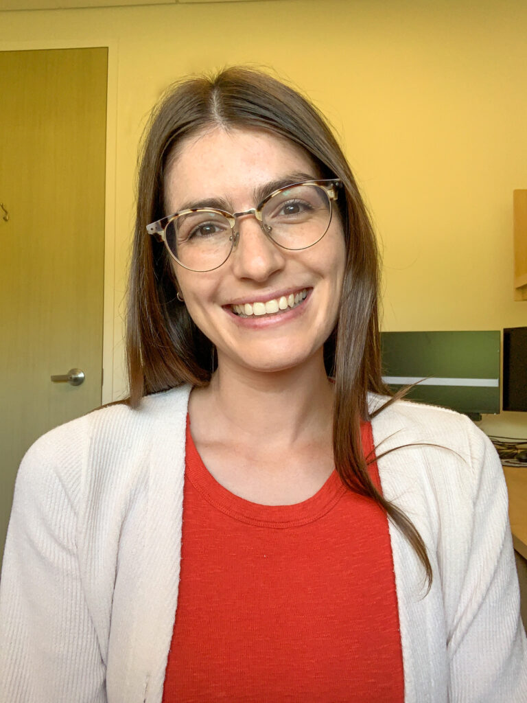
Adi Foord is a Scialog Early Science with LSST Fellow. (courtesy of Foord)
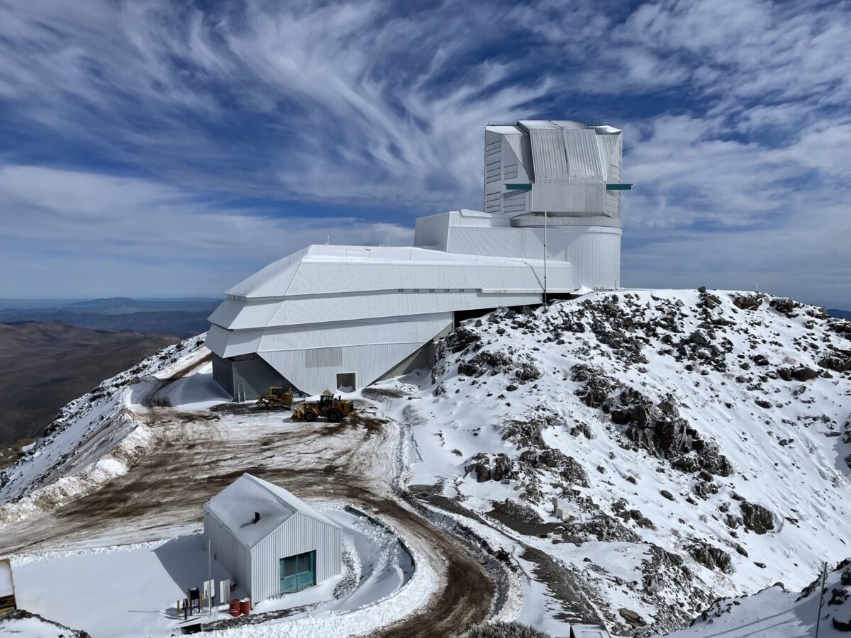
In this role, Foord has the opportunity to propose research analyzing data coming from the Vera C. Rubin Observatory, the largest ground-based telescope in the world. Over the next 10 years, it will carry out the Legacy Survey of Space and Time (LSST), scanning the entire sky every three days. Construction of the observatory, in Chile, is nearly complete, and its first images of the sky are expected later this year. LSST will collect an unprecedented amount of astronomical data ripe for analysis—and full of discoveries just waiting to be uncovered.
The Scialog fellowship is designed to hasten those discoveries. A cohort of 50 fellows includes early-career researchers in a wide range of fields: astrophysicists like Foord, theoretical physicists, data scientists, software engineers, and more. Each year of the three-year fellowship, the fellows gather for a multi-day workshop designed to facilitate development of innovative research proposals that would use LSST data.
Foord’s first proposal, in collaboration with Krista Lynne Smith at Texas A&M University, is one of eight proposals selected for funding out of the 33 submitted after the first Scialog workshop.
Below, Foord answers questions about the fellowship, her research at UMBC, and her passion for physics, and shares advice for aspiring astronomers.
Q: How is this fellowship unique?
A: LSST’s open-access dataset will be higher in volume and complexity than any previous astrophysics survey and will contain unexpected data that will provide insights into fundamental questions about the universe.
This fellowship addresses two key challenges to unlocking the discoveries in LSST data: 1) the lack of seed funding to support early LSST discoveries, and 2) the need to create cross-disciplinary connections to address the ambitious questions LSST is poised to answer. By facilitating creative proposals from interdisciplinary teams and then funding those proposals, the fellowship program addresses both of these challenges.
On top of this, I was really excited by the interdisciplinary nature of the program. The most creative and innovative ideas often emerge when scientists from diverse fields come together. By fostering collaboration—over food, coffee, and discussions—the fellowship provides an ideal environment for sparking ideas.
Q: What are the goals of your funded proposal?
A: My proposal is focused on building and using a “Dual Active Galactic Nuclei (AGN) Finder” on LSST data. Dual AGN are pairs of actively accreting supermassive black holes whose galaxies are merging. They are very difficult to find, because they generally just look like one source, instead of two.
Thankfully, the brightness of different kinds of objects in the sky varies predictably over time, so a graph called a “lightcurve” that represents an object’s varying brightness can be used to identify an unknown object. Our tool will use the unique lightcurve expected from dual AGN systems to search for pairs of merging AGN. As of right now, there are less than 100 confirmed pairs of merging AGN, so we hope to dramatically expand this sample size using our tool. LSST scans the full sky every three days, so it will have millions of lightcurves of AGN. It’s the perfect observatory to carry out such an analysis.
This new project with LSST nicely bridges my Ph.D. work on dual AGN with my current interests in how galaxy mergers influence supermassive black hole growth. It also opens the door to utilizing a completely different type of data.
Snowfall blanketed the mountains of northern Chile on August 4, 2024, including Cerro Pachón, where Rubin Observatory is located. (NOIRLab/NSF/AURA/F. Bruno)
Q: Why are you passionate about this field, and how did you find your way to it?
A: I wasn’t sure what I wanted to pursue in college until my senior year in high school, when I had the opportunity to take an observational astronomy class. Prior to that, I enjoyed physics and math, but I hadn’t found the applications particularly exciting. That all changed the first time I looked through a telescope and held a meteorite that had originated from a collision on Earth about 50,000 years ago. The sheer excitement I felt in those moments made me realize that astrophysics was the perfect intersection of math, physics, and an interesting environment. The support I received from my high school physics (Mr. Woosnam) and astronomy (Mr. P) teachers gave me the confidence to pursue this path.
I still experience that thrill every time I open new data, not knowing what discoveries await, but eagerly anticipating what the data will reveal. The unwavering support of my mentors and friends has enabled me to stay the course and be successful in this field. Now, as a professor, I strive to inspire that same excitement in my students and help them discover their own passions.
Q: What do you enjoy about being a member of the physics department and the UMBC community more broadly?
A: Since joining UMBC in fall 2023, I have been truly impressed by the students. The undergraduates are incredibly passionate about joining research teams and learning about astrophysics. It has been a rewarding experience to start building my own research group of graduate students and mentor them through their first graduate research projects.
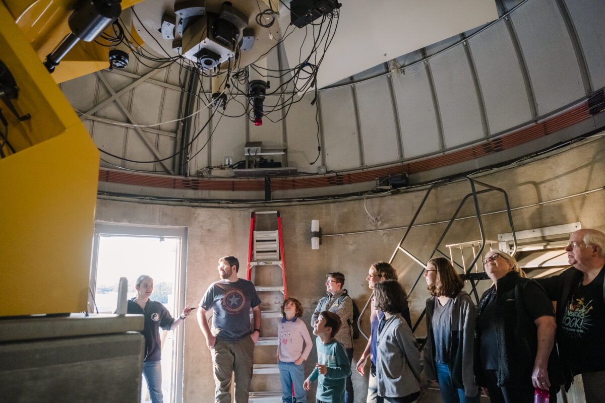
A UMBC physics student gives a tour of the observatory on top of the Physics Building to members of the local community. (Marlayna Demond ’11/UMBC)
I also particularly appreciate the amount of resources available for new faculty. In my first year, I participated in three different programs that introduced me to faculty across various departments, which I found invaluable. The Eminent Scholar Mentoring Program is especially unique—it has allowed me to professionally and personally connect with a seminal scientist in the field, someone I may not have had the opportunity to easily meet otherwise. These resources truly set you up for success and make you feel incredibly supported.
Q: What advice do you have for aspiring astronomers?
A: At the undergraduate level, actively seek out research opportunities wherever you can. Look for options at your own institution, nearby universities, and through programs offered by various agencies. Even if the research isn’t in an area you originally wanted to pursue, these experiences will help you develop critical thinking, problem-solving skills, and the ability to ask important questions.
I also encourage you to surround yourself with supportive mentors and colleagues. Every successful scientist got to where they are because they had people backing them along the way—the “lone genius” is rarely the full story. Being part of a supportive community not only helps you move forward but also fosters creativity and passion in your research, which is ultimately what drives us.
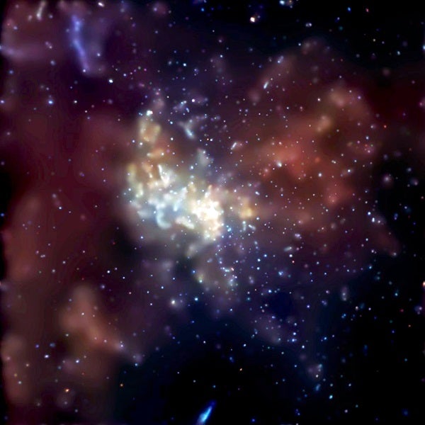
Sagittarius A*, the black hole at the center of our Milky Way galaxy, is located at the brightest, central point in this image. (NASA/CXC/MIT/Frederick K. Baganoff, et al.)
Q: Anything else you are burning to share?
A: My favorite supermassive black hole, of course! It’s Sagittarius A*, the supermassive black hole at the center of our Milky Way galaxy. While it’s puny (only about four million times the mass of our Sun, which is relatively small for a supermassive black hole) and not particularly active, its proximity to us makes it an incredibly intriguing object. Despite being so close, there are still many unanswered questions about its growth and activity. Most of my research focuses on supermassive black holes in other galaxies, but if I had unlimited time, I would love to dedicate more of my work to studying Sagittarius A*.
A new course in UMBC’s Department of Mathematics and Statistics is having a positive impact on student success in a notoriously difficult course for math majors everywhere. Two new papers by UMBC mathematicians and members of UMBC’s Faculty Development Center strongly suggest that MATH 300: Introduction to Mathematical Reasoning(IMR) is helping students succeed in MATH 301: Real Analysis, the first course math majors take that relies on one’s ability to construct and analyze proofs, rather than just do calculations.
In Real Analysis, “We’re switching gears of how students think. They go from calculational things to proof-based work,” says Kathleen Hoffman, professor of mathematics and lead author on the new papers. “Now your solution is a paragraph that you have to write in full sentences. It has to have logical structure. It has to start with a hypothesis and end with a conclusion. It’s a big hump for students to get over.”
Real Analysis is easily one of the most challenging courses for math majors nationwide, Hoffman says. Over the years, many institutions have introduced a preparatory course that teaches students how to develop proofs without requiring them to learn new math content at the same time. The conventional wisdom is that these courses help, but almost no one had conducted a rigorous study to find out.
“There was a huge gap in the literature,” Hoffman says.
Forming the team
UMBC math faculty had seen the need and been talking about adding a dedicated proof-writing course for years, but it hadn’t quite come together. Hoffman jump-started the process by applying for a Hrabowski Innovation Fund Grant in the Scholarship of Teaching and Learning category. These awards support faculty who want to do ambitious projects that they might not otherwise have bandwidth for.
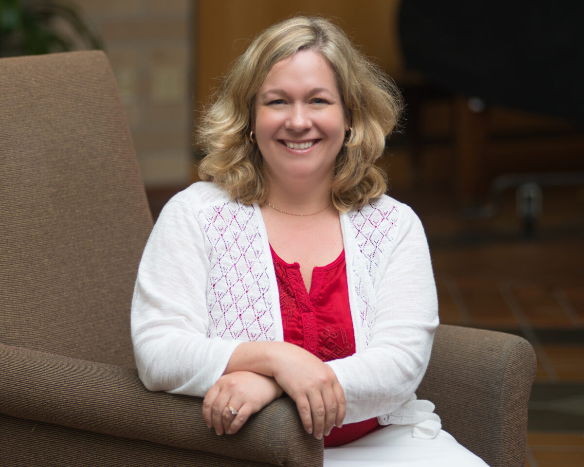
Kathleen Hoffman wrote the Hrabowski Fund for Innovation proposal that supported the team’s efforts to rigorously evaluate the effectiveness of their new course. (Courtesy of Hoffman)
When the award was funded, Hoffman formed a team with Justin Webster, associate professor of mathematics, and Kal Nanes, associate teaching professor of mathematics, to design the course for UMBC. Webster had re-designed and updated one of these proof-writing courses at his previous institution, the College of Charleston. The math team also worked with staff in UMBC’s Faculty Development Center to design a rigorous study to evaluate the course’s effectiveness over time. The team knows of only one other such study, from the 1980s, despite the rising incidence of proof-writing courses at universities nationwide.
Hoffman, Webster, and others believed that IMR would help UMBC math students, and informal observations supported their hunch once the course launched. These publications provide statistical analyses to back their intuition, and now Retrievers and students from other institutions can benefit from their successful formula.
Thinking about thinking
The first paper, published in a special issue of Educational Sciences, focused on written reflections the students completed every week along with their proofs. Prior research suggests that students tend to struggle in specific skills related to proof-writing, so the students were required to address how well they thought they did on each of four skills in their reflections.
“Students who did very thoughtful responses did much better in this course, but they also did much better in Real Analysis, where they didn’t do any reflections,” Hoffman says. The reflections “give the students a framework for understanding what they know and what they don’t know. It gives them the words to use.”
The study analyzed the quality of the reflections, but not necessarily the content. It didn’t seem to matter exactly what aspects of proof-writing the students addressed in their writing—simply the act of metacognition, or “thinking about thinking,” seemed beneficial.
The correlation was strong, but Hoffman admits the study does not prove causation. Strengthening the evidence, though, is that thoughtful reflections in IMR were not correlated with success in its prerequisite course, MATH 221: Introduction to Linear Algebra. That suggests the reflections, and potentially other elements of IMR, were the difference-maker for students moving forward.
A solid foundation
A second paper, published in the International Journal of Mathematical Education in Science and Technology, compared students’ grades in Real Analysis depending on whether or not they had taken the proof-writing course. The findings showed that IMR did not much affect the outcomes for students who earned As in the prerequisite linear algebra course—they were also likely to do well in Real Analysis whether they took IMR or not. However, students who earned a B or C in the prerequisite course were much more likely to successfully complete Real Analysis if they had taken IMR.
The researchers also received overwhelmingly positive feedback from students who had taken IMR about its benefits. One student said,
“I feel like [IMR] gave me a solid foundation in understanding how to write proofs, which allowed me to come into [Real Analysis] with a bit more confidence. Without it, I probably would have struggled through [Real Analysis] since I would have been learning how to write proofs and the [Real Analysis] material at the same time.”
Based on the results of these studies, the UMBC mathematics and statistics department has decided to make IMR a required part of the curriculum for math majors and minors. Minors used to take Real Analysis as their terminal course, but now they take IMR. For majors, IMR provides the foundation needed to support success in Real Analysis.
More than pushing symbols around
When he took it as an undergraduate, an IMR-type course “was the thing that made me want to be a math major,” Webster says, so designing this course for UMBC was an exciting prospect. Becoming proficient in writing and analyzing proofs, rather than doing calculations, is like “writing versus writing literature,” he says—you have to spend a lot of time thinking about what you’re trying to accomplish and how to structure your arguments. You can’t just “plug-and-chug,” applying various theorems and techniques to instances of a given type of problem. “Math isn’t just the act of pushing symbols around,” Webster says.
Justin Webster often meets with students to support their progress in math courses. (Marlayna Demond ’11/UMBC)
To prepare students to write mathematical literature, Hoffman says that in IMR, “In one sense I’m teaching math, but in another sense I’m not. I’m teaching them how to think—how to structure their argument and express it clearly.” Almost never can a student simply sit down and write a proof in one sitting, like completing a problem set in prior math courses. It’s more like writing a paper.
When you work on a proof, “You think, you don’t get it, you go do something else, you think, ‘Oh, I think I know what to do,’ you come back, and that is normal,” Hoffman says. “They have to understand, this is not instant gratification—you will struggle with this and I’m expecting you to. It’s inevitable that they will struggle—I’m teaching them to persist through the struggle.”
A collective commitment
The studies would not have been possible without support from the Faculty Development Center. While many faculty might like to conduct more rigorous analysis of their teaching methods, it’s not their area of expertise. “If you want people like me who do disciplinary research to engage in pedagogical research, you have to give me some help,” as Hoffman put it.
Kerrie Kephart was one of several members of the Faculty Development Center who contributed to the study of the new course’s impact. (Marlayna Demond ’11/UMBC)
That’s where the FDC came in. Several staff members became involved in the project, including Tory Williams, Jennifer Harrison, Kerrie Kephart, and Linda Hodges, adding their individual areas of expertise.
“The math faculty have deep expertise in math pedagogy, but needed our support to help plan the intervention, design the research study, and analyze the data. The project required all hands on deck.” shares Kephart, co-author on the written reflections study and interim FDC director. “As a qualitative researcher with expertise in the teaching of academic writing, I enjoyed the challenge of figuring out how to study the effects of incorporating reflective writing into a math class.”
The project is also a demonstration of the math department’s commitment to supporting student success, even if that required some culture change. Since IMR’s initial offering in 2019, several additional math faculty have taken on teaching the course. Each time someone new takes it on, they work closely with experienced instructors, and all sections of the course are closely coordinated to ensure quality and consistency for students.
The project is a masterclass in recognizing a challenge (a high failure rate in Real Analysis) and taking creative, concerted, and collective action to address it, with very positive results. “After realizing there was a gap in students’ preparation, a group worked together to fill it, and in the process learned a lot about how to measure progress in math education and pedagogy,” Webster says. “We leaped at this opportunity to effect change and measure outcomes in a novel and modern way.”
And because they carefully evaluated the project’s effects and published the results, now other math departments can benefit from their findings. That possibility was a highlight for Kephart. “Since our work in the FDC generally supports faculty, teaching, and learning here at UMBC,” she says, “it’s exciting to make a contribution toward the development of math pedagogy beyond our campus.”
UMBC has received $1.5 million from the National Institute of Standards and Technology to organize a new UMBC Quantum Science Institute. The funding will support graduate fellowships for students pursuing quantum technology research, the development of new courses and academic programs focused on quantum, and equipment to enhance existing quantum labs and start new ones.
“This institute is the crowning jewel of several decades of pioneering quantum research here at UMBC,” says QSI director and professor of physics Todd Pittman, Ph.D. ’96, physics. “We have some heavyhitters here who are founders in the field, and the QSI is building on that foundation with a new generation of outstanding quantum-focused faculty.”
Quantum technologies harness the odd behaviors of particles at the atomic level to generate new functions, and are typically much more powerful than their conventional technology counterparts. To approach quantum research from every angle, the institute includes faculty across the College of Natural and Mathematical Sciences and the College of Engineering and IT in physics, mathematics and statistics, computer science, and information systems. They are pursuing quantum research in areas including computing, communications, sensing, information theory, algorithms, and more. “You just can’t do comprehensive quantum research without bringing together engineers, data analysts, computer scientists, mathematicians, and physicists,” Pittman says.
“I am thankful for the support provided by NIST to develop the UMBC Quantum Science Institute under the leadership of Dr. Pittman,” shares Karl V. Steiner, vice president for research and creative achievement. “QSI will catalyze a new quantum research effort, in support of Governor Moore’s ‘Capital of Quantum’ Initiative to position Maryland as a global leader in this rapidly growing field.”
Training quantum-ready workers
In addition to contributing to research innovation and discovery, the institute will train students from a range of backgrounds to take on skilled roles in the booming quantum industry.
“The quantum industry has exploded,” Pittman says. The quantum technology market was valued at $10 billion in 2021 and is projected to rise to $44 billion by 2028, with potential for much more growth beyond that. In addition to tech giants like Google and IBM, hundreds of start-ups are working in the quantum space. “The market is hungry for quantum-ready workers,” Pittman says, and UMBC is ready to train them.
UMBC marked World Quantum Day in 2022 with a short video highlighting the quantum technology research happening in the physics department.
Sandra Cheng, a fourth-year Ph.D. candidate in physics, is a member of the first cohort of quantum graduate fellows. “Quantum science research, particularly in computing and networking, is quite interdisciplinary by nature,” she says, “and I’m hopeful the QSI will bring together like minds from all the departments involved, so that we’ll be able to contribute towards a greater understanding of quantum science together.”
The QSI leadership team plans to offer social and professional development programming for the fellows. In addition to preparing them for the workforce, the connections they make will help generate a personal network of support, encouraging persistence in a demanding field. Organizing interdisciplinary offerings for the student cohort will also promote collaboration among faculty, Pittman says, adding, “Students are the glue that holds the center together.”
“This new institute is a celebration of our strength and history in quantum research,” Pittman says. “I’m looking forward to seeing QSI unify the quantum researchers on campus in a way that promotes and facilitates interdisciplinary collaboration.”
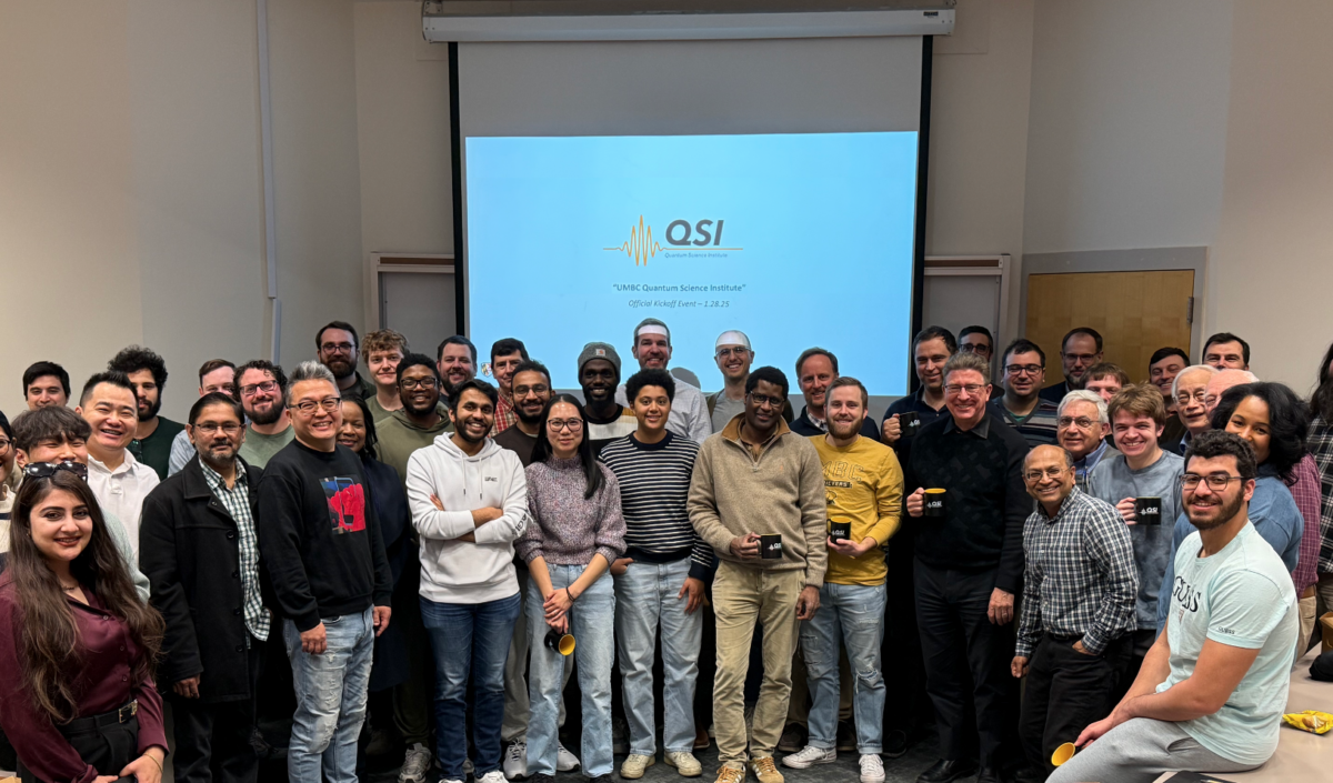
Representatives from all of the departments involved in the new QSI recently attended a launch event.
Some mathematical and statistical challenges are so fundamental that the solutions can be applied to an array of real-world applications—which we all benefit from. But it’s not necessarily the applications that excite researchers. They’re on the hunt to develop an elegant set of equations or a more accurate model because of the work’s inherent beauty and satisfaction, and, simply, because it’s fun.
This past fall, members of the UMBC Department of Mathematics and Statistics received at least three major grants to pursue work with significant real-world potential, from self-driving cars to treatments for Alzheimer’s. And while the mathematical work underpinning these potential uses can seem opaque, abstract, and just plain hard to many people, these mathematicians and statisticians see the process of thinking about the challenges as exciting and stimulating in and of themselves.
Optimizing optimization
Andrei Draganescu (courtesy of Draganescu)
Andrei Draganescu, associate professor of mathematics, has received a grant from the National Science Foundation to pursue a creative new approach for solving optimization problems. Anytime a system performs a basic functionality, but you would like to make it better or more efficient, that’s an optimization problem, Draganescu explains.
Optimization means maximizing or minimizing something, such as cost, time, or power output. Think about today’s large language models, such as ChatGPT: Before it can respond effectively to any prompts, the model must rely on an algorithm to “learn” an optimal set of parameters from a vast amount of training data. Only once those parameters are fixed can it use them as a basis to determine the best response to your prompt. The time, electricity, and human work hours involved in training can cost billions of dollars, not including the capital investment in the hardware itself—so even a very small increase in efficiency makes a huge difference.
All of the possible combinations of parameters can be represented as points along a curved surface with hills and valleys. An algorithm developed by a mathematician can search for the lowest or highest point on that surface to solve the optimization problem, depending if you are looking for a minimum or a maximum. One risk with these algorithms is that they may end up at a local minimum or maximum—the peak of a single hill or the base of a single valley—that isn’t actually the global maximum or minimum across the entire surface.
In his grant application, Draganescu proposed a novel way of solving this kind of problem. “Because this type of problem is so old, it’s hard to find something new. But I think I did,” Draganescu says. Rather than searching for the minimum or maximum along a series of straight lines that approximate its curved surface, Draganescu has proposed a way to search directly along a set of well defined curves. The idea for the new approach “is a completely new angle,” he says, and in correspondence with colleagues to date, “nobody has contradicted me so far.”
All of the possible combinations of parameters for a system can be represented as points along a curved surface with hills and valleys. (Fiona Suherman/UMBC)
Sticking with the hard problems
In math, there are many problems that are easy to express but extremely difficult to solve. Some have puzzled mathematicians for centuries. Draganescu’s Ph.D. advisor encouraged his students to think about these problems from time to time, in case they found a solution, of course, but also to keep their creative juices flowing. Draganescu continues the practice today.
“Sometimes I’ve spent days in a row on some of these very hard problems that nobody knows how to solve,” he says. “And of course I would love the glory, but honestly, that’s not where the fun is. I’m old enough to know that the fun is actually thinking about it.”
“And of course I would love the glory, but honestly, that’s not where the fun is. I’m old enough to know that the fun is actually thinking about it.”
Andrei Draganescu
Draganescu’s sticktoitiveness sometimes pays off in his everyday work. In the case of the idea that underpins his recent proposal, “I was surprised that it actually worked out. But ‘working out’ means many many months of work,” he says. “From having the initial idea to trusting that it could go somewhere, there were a lot of mistakes in the calculations and all that.” So why did he persist, knowing there was a good chance it wouldn’t “work out”? “I stuck with it because it was fun. I enjoy thinking about these problems.”
Draganescu is continuing to develop his new technique for solving optimization problems, which could improve efforts to optimize all kinds of systems, from AI to airline schedules to agriculture—all because he found joy from sitting with a sticky problem.
Replicating the real world
Ansu Chatterjee (courtesy of Chatterjee)
Ansu Chatterjee, professor of statistics, is another UMBC math mind whose work could have a major impact on a wide range of fields. He and UMBC colleagues Animikh Biswas, professor of mathematics, and Karuna Joshi, professor of information systems, have received an NSF grant to expand the use of “digital twin” technology. The grant proposal highlighted the potential to advance diagnosis and treatment for neurodegenerative diseases such as Alzheimer’s or Parkinson’s, but digital twin technology has a huge range of potential applications.
A digital twin is “essentially a clone of something in the physical world replicated inside a computer,” Chatterjee says. Additional applications could include forest management or weather forecasting. These twins can offer up predictions about which direction a hurricane will turn, which treatment protocol might be most effective for a particular patient, or whether it’s the right time for a prescribed burn in a forest—but there will always be uncertainties in the models.
“Measuring those uncertainties is a huge task,” Chatterjee says. How to deal with those uncertainties is one of the things the team wants to focus on with the new grant, which is very statistics-heavy, Chatterjee notes. Understanding and reducing the uncertainties requires understanding the physical processes at play in the real-world version of the system, and then working them into the model—and that’s hard.
Beyond the black box
Typical AI models are frequently “black box models,” which means even the people who created them don’t always understand exactly what they’re doing. For one thing, they are not constrained by objective truth in the physical world, Chatterjee explains. “It’s not even attempting to get the biology or the chemistry correct,” he says, and that’s not acceptable for a digital twin that’s modeling the human brain and may be used to determine a real human’s treatment plan.
“You don’t get the model right unless you get the underlying math of the molecules that constitute it right. So mathematics and statistics have to be the underpinning of anything related to digital twins.”
Ansu Chatterjee
“You don’t get the model right unless you get the underlying math of the molecules that constitute it right,” Chatterjee says. “So mathematics and statistics have to be the underpinning of anything related to digital twins.”
The digital twins Chatterjee and colleagues hope to further with their new research need not only replicate the outcomes of a system, but also the system’s mechanisms and internal processes. Again, that’s hard—especially when humans don’t yet fully understand many of these systems, such as the human brain, how cancer develops, or how various environmental factors influence carbon sequestration, for example.
“We are at the foundation of digital twins,” Chaterjee says. Once they are developed to a point where they are usable, digital twins and AI more generally would “open up a vast amount of opportunities for making fairly rapid progress with the actual science. This is where AI can help in scientific progress. It can suggest opportunities and possibilities which then can be verified,” Chatterjee says. “It would rule out certain possibilities. There would of course be false positives and false negatives, but as digital twins keep improving, it would be more and more convenient to use those first to find out a lot more interesting things.”
A digital twin is “essentially a clone of something in the physical world replicated inside a computer,” Ansu Chatterjee says. (Illustration by Sadaf Rehman ’19/UMBC)
Fundamental building blocks
Chatterjee and colleagues are aware of the risks associated with digital twins. “There’s quite a lot of ethical issues that are related to digital twins in general, and it goes up several notches when it’s digital twins related to humans.” For example, there are regulatory concerns around who owns the data put into a digital twin (such as someone’s genomic data) and who is compensated when the twin is used for profit. “It’s exciting, and it’s also kind of a nightmare,” Chatterjee says.
That’s why he and colleagues will be working directly with federal regulatory agencies to make recommendations about how to handle these AI models. “This grant is sort of laying down the first fundamental steps—saying this is what should be done, this is how the first steps should be done,” Chatterjee says.
While that work is critical, it’s the math that truly excites Chatterjee. “For me, it’s about building the fundamental building blocks, whether it’s cancer or neurodegenerative diseases or forests” he says. “The essential ingredients for the digital component of it rely on the same math, the same statistics.”
Defining possible
Matthew Kvalheim (courtesy of Kvalheim)
Matthew Kvalheim, assistant professor of mathematics, is also working on developing tools that can help people in a wide range of situations. His ultimate goal? To be able to tell researchers whether whatever they are trying to do with their system is possible or impossible. Sound vague? That’s the point.
“I’m never thinking about a specific application,” Kvalheim says. “I’m always working at the fundamental level, with a class of mathematical models that can describe all of them.” For Kvalheim, it’s the underlying math that is exciting—which is fortunate for all the people working on applications who will eventually use his multi-purpose tools.
“For any real world system, you might want it to do some behavior, or not do some behavior,” Kvalheim says. This might be making sure two self-driving cars don’t collide, preventing a humanoid robot from falling on its face, or keeping an electronic device at a safe temperature.
“Lots of people work on the set of models that I study,” he says, and the vast majority of them are investigating a particular application. These other researchers are very good at coming up with solutions that result in stability, or maintaining a certain state in a system, and safety, or preventing dangerous states in the system, “but there’s one thing they can’t do,” Kvalheim says. “They can’t determine if the thing they are trying to do, to make their system stable and safe in the way they want, is just fundamentally impossible. There could be a fundamental law of nature that says too bad, you can’t do it. You could try forever, but the heat death of the universe will occur first.”
A tool Kvalheim has already published to help prove impossibility relies on math that is closely related to something called the “hairy ball theorem.” If you imagine certain 3D shapes covered by directional vectors—or, to make it simpler, hairs—”Try as you might, you can’t comb that hair flat everywhere without creating a cowlick somewhere,” Kvalheim says. (Illustration by Sadaf Rehman ’19/UMBC)
A license to look
Kvalheim would like to discover new fundamental limitations that hold true for any application you could come up with, and also new fundamental capabilities. The U.S. Air Force recently awarded him a grant to further this work. He’s trying to generate a sort of mathematical litmus test that any practitioner can use to find out whether the thing they are trying to do with their system (whatever it is!) is fundamentally possible or impossible. If it’s impossible, it saves them a lot of time, energy, and money. And if it’s possible? “It’s like giving people a license to go look for it,” Kvalheim says.
The best part is that the end user of one of Kvalheim’s tools wouldn’t need to understand all the pure math behind it. “The idea is to produce useful tools that they can use even if they don’t have the time or interest in diving into all the details,” he says.
Kvalheim’s stability research is applicable even to the Lorenz system, which is a set of ordinary differential equations. Solutions to this system can vary chaotically depending on the initial parameters, but converge toward and stay within a bounded space. This space appears butterfly-shaped when represented visually. (Illustration by Fiona Suherman/UMBC)
Math undergirds just about everything, Kvalhiem says, so it’s important to study the general case, rather than focus on specific applications. He wants people to see that it’s important “to know when there are fundamental laws of nature telling them that it is impossible or possible to do things that they want, and that math allows you to discover such laws of nature—and not just for one real-world system, but for many systems all at once.”
Somewhere a math or stat researcher is helping prevent your car from crashing, providing life-saving medical tools for yourself or a loved one, reducing wildfire risk where you live, or improving your experience with an AI tutor—and you might never even know it. There are humans behind each of these innovations who find joy and value in the math puzzles and problems that are all around us. And some of them work at UMBC.
Carla Guarraia, Ph.D. ’06, molecular and cellular biology, has received a Presidential Award for Excellence in Mathematics and Science Teaching for her work at the Park School in Baltimore. Only one award is given per state per year, and Maryland is an extremely competitive region.
In 2017, Guarraia proposed and coordinated a complete redesign of the science curriculum for ninth and 10th graders at Park in close collaboration with colleagues. The new ninth grade course, first implemented in 2019, integrates physics, engineering, and computer science content, and the 10th grade course brings together concepts from biology, chemistry, and data analysis. In addition to covering content, the courses emphasize skill-building and opportunities for memorable and authentic project-based experiences that inspire excitement about science. Park juniors and seniors are then prepared to pursue an array of electives in all five disciplines and interdisciplinary topics.
An external evaluator lauded the resulting curriculum, which Park calls its “Core Program,” for its alignment with the Next Generation Science Standards, differentiated learning approach for students with a range of needs, emphasis on mastery learning and a growth mindset, prioritization of active learning, and the faculty’s commitment to caring for their students.
Expanding her view
Guarraia, who as of the 2024 – 2025 school year is the upper school science department chair at Park, studied for her Ph.D. under the mentorship of Philip Farabaugh, professor of biological sciences. Then she completed a postdoctoral fellowship with Jennie Leach, associate professor of chemical, biochemical, and environmental engineering.
“Phil Farabaugh supported my growth as an academic, and as a citizen of the world. He consistently showed respect for me and built up my confidence that I could be smart and fun while not only teaching science but caring for the people around me. As a mentor for life, I have learned from Phil that if I am my authentic self my life will be most successful,” Guarraia says.
Leach “shares my passion for teaching, and in Jennie I continue to have a role model for pursuing ambitious exciting science while also being a working mom,” Guarraia adds.
“UMBC is a really special place that shaped my view of Baltimore, as the campus focus was on rigorous education, inclusion, and excellence, and it was my first experience with a truly international community,” Guarraia says. “I am forever grateful for how UMBC, Jennie, and Phil expanded my view of the world and of myself.”
A large international team of scientists has observed a phenomenon that astronomers didn’t ever expect to see happen in real time. A new paper published in Astrophysical Journal Letters led by Eileen Meyer, associate professor of physics at UMBC, describes the findings. It reports remarkable increases in radio emission in a few months and formation of plasma jets extending from a black hole over the course of a year.
A galaxy about 270 million light-years away from Earth in the constellation Draco called 1ES 1927+654 is the focus of the excitement. For many years, scientists had classified 1ES 1927+654 as an “active galactic nucleus,” or AGN, meaning it has an active black hole at its center. This particular black hole was adding material at a slow rate—until it wasn’t.
Back in 2018, the black hole first made news when it suddenly increased its activity exponentially. It dramatically increased the rate at which it was consuming material and became over 100 times brighter in the visible light spectrum over the course of a few months. A shift like that was once thought to take far longer than a human lifetime, on the order of thousands to millions of years. Since then, scientists have been observing it closely for any additional interesting phenomena, and 1ES 1927+654 has delivered.
More drama
After the major increase in activity began in 2018, which included nearly a year of extremely high levels of X-ray emission, the black hole quieted down again by 2020—only to dramatically increase its output again in 2023. At that time, it began emitting radio waves at 60 times the previous intensity over just a few months, behavior which has never been monitored in real time for a supermassive black hole.
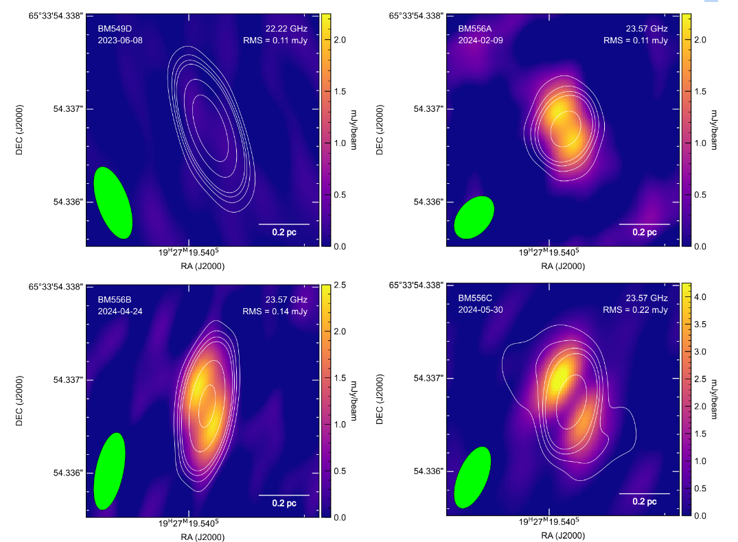
Some of the highest-resolution imaging of radio frequency emissions was collected using a technique called Very Long Baseline Interferometry (VLBI). It clearly shows a pair of oppositely directed plasma jets forming near the black hole and expanding outward over the course of 2023 – 2024. Among the other unusual behavior of the black hole, this is the first-ever observation of jet formation in real time.
In recent years, scientists have discovered a handful of supermassive black holes that appear to emit far more intensely at radio frequencies compared to when they were first observed, which they call “changing-look AGN.” However, until now all of them had been observed at two timepoints years or decades apart, and the assumption was that “something happened” in between. This new paper gives the very first look at how this kind of change occurs in detail.
This figure from the new paper shows the plasma jets (yellow blobs) forming. Clockwise from top left, the dates of the images are June 2023, February 2024, April 2024, and May 2024.
Turning on in real time
In some cases, black hole jets “can reach huge scales well outside the host galaxy. They can affect how many stars are forming,” Meyer says. Figuring out how the jets work “is a very important thing, in order to understand the big picture of how the universe is evolving and galaxies evolved.”
Sibasish Laha (courtesy of Laha)
In the case described in the new paper, “We have very detailed observations of a radio jet ‘turning on’ in real time, and even more exciting are the VLBI observations, which clearly show these plasma blobs moving out from the black hole,” Meyer says. “That shows us that this really is an outflow jet of plasma that’s causing the radio flare. It’s not some other process causing increased radio emission. This is a jet moving at likely 20 to 30 percent of the speed of light originating very near a black hole. That’s the exciting thing.”
Sibasish Laha, an associate research scientist for UMBC with the Center for Space Sciences Technology at Goddard Space Flight Center and second author on the new paper, has long studied changing-look AGN at X-ray wavelengths. On a hunch that 1ES 1927+654’s radio frequency emission might show interesting behavior as well, he reached out to Meyer to form a collaboration to study it and other similar galaxies back in 2020. He is lead author on a companion paper that is currently under review. It includes additional X-ray observations and interpretation of the jet formation event.
“We still do not understand how black holes and their host galaxies interact with each other and co-evolve in cosmic time,” Laha says, “and this study for the first time gives us the rare opportunity to understand how a supermassive black hole ‘talks’ to the host galaxy.”
Not for the faint of heart
In this kind of work, time is of the essence. “Time-domain astronomy,” as it’s called, “is not for the faint of heart,” Meyer says. “You know, there are rapid alerts—something happens and you have to go follow up. You gotta get on it, and it doesn’t matter if it’s midnight, you have to send that email because you know every hour counts. It’s a little stressful.”
Onic Shuvo (courtesy of Shuvo)
The project became an “all hands on deck” moment for the UMBC collaboration. Once Meyer and Laha saw the huge jump in radio activity in 2023, Meyer says, “We were like, ‘whoa, ok, something is happening.’ This has never been seen before. We got very excited, so this is where we went all in on basically trying to grab every radio telescope and get it to look at this source.”
Because 1ES 1927+654 was changing so rapidly before their eyes, the team was awarded new, unscheduled observations on telescopes around the world during the study period, when typically telescope time must be scheduled months or years in advance.
A postdoctoral fellow working with Meyer, Onic Shuvo, who is third author on the paper, took on the lion’s share of the late-night duties, rapidly analyzing incoming data and requesting new observations. He’s thrilled to be part of such an exciting discovery. “This remarkable finding challenges existing models of AGN activity and highlights the unique role that changing-look AGN play in unraveling the mysteries of the central engine of active galaxies in real-time,” Shuvo says.
A new jet is born
The newborn jets coming from 1ES 1927+654 are relatively small compared to the massive jet structures in some of the most powerful AGN, Meyer says. But that doesn’t make them less interesting—in fact, they are probably more common across the universe and therefore very important to understand, she says.
Some data suggested that the 2018 flare, in the visible spectrum, could be due to a “tidal disruption event,” where a large object like a star or cloud of gas gets too close to an inactive black hole and artificially brightens it for just a few years, Meyer says. But observations of tidal disruption events in already-active galaxies are rare and not well understood.
While the largest plasma jets extend well beyond their host galaxies and last millions of years, scientists are gaining understanding of a new class of smaller, shorter-lived jets called “compact symmetric objects,” or CSOs. Meyer believes the data in this case point most strongly to the birth of a new CSO. One recent hypothesis is that jets in CSOs are qualitatively different from the very large and long-lived jets seen elsewhere, Meyer says, perhaps representing “a single ingestion of a star or a gas cloud; basically a single tidal disruption event happens and powers this short-term jet for maybe 1,000 years.”
Perhaps the tidal disruption event occurred several years ago, “and it took a few years for the accreting black hole to organize and start producing the jet,” as the team saw in 2023 – 2024, Meyer says.
Eileen Meyer works with students in her laboratory. (Marlayna Demond ’11/UMBC)
Open questions
Overall, “We still don’t really understand after all these decades of studying these sources why only a fraction of accreting black holes produce jets and then exactly how they launch them. Until recently we could not literally look into that innermost region to see what’s happening—how the accretion disk surrounding the black hole is interacting with and producing the jet. And so there are still a lot of open questions there,” Meyer says.
Questions remain, but today there are many promising models of how black holes produce jets, Meyer says. Next steps will include working with theorists to understand how the data from this study can help test and refine those models.
“There’s a lot of theoretical work to be done to understand what we’ve seen, but the good thing is that we have a massive amount of data,” Meyer says. “We’re going to keep following this source, and it’s going to continue to be exciting.”
In fall 2022, the College of Natural and Mathematical Sciences at UMBC and the University of Maryland School of Medicine (UMSOM) received a five-year, $13.7 million grant from the National Institutes of Health (NIH) to enhance recruitment and training of junior faculty and promote inclusive excellence at UMBC. Since then, UMBC and UMSOM teams have been planning programming for the incoming FIRST (Faculty Institutional Recruitment for Sustainable Transformation) cohort, undertaking faculty searches, and otherwise preparing for their new colleagues’ arrival.
As of this fall, four new UMBC faculty members and three new UMSOM faculty have been selected, with three more to be added at UMSOM. Two of UMBC’s hires are busy doing research, recruiting students, and settling into their new positions. The other two will officially assume their roles in January, but they are already visiting campus and starting to set up their laboratory spaces.
In November, the leads on the grant from both institutions and all the new hires gathered at UMBC for a reception to celebrate their first semester and build community among the cohort.
A talented group
“Although I have only been on board for three months, I already feel the leadership’s dedication to supporting early-career faculty, particularly those from underrepresented backgrounds, through professional development and tailored resources,” shared Cheng-Yu Li, a new assistant professor of biological sciences at UMBC hired through the program. “I am excited to work alongside this talented group of scientists and contribute to advancing neuroscience research, education, and outreach.”
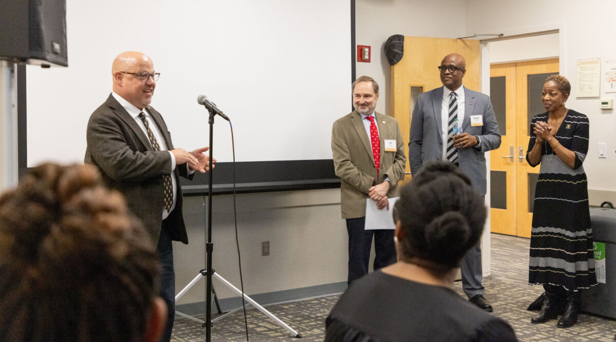
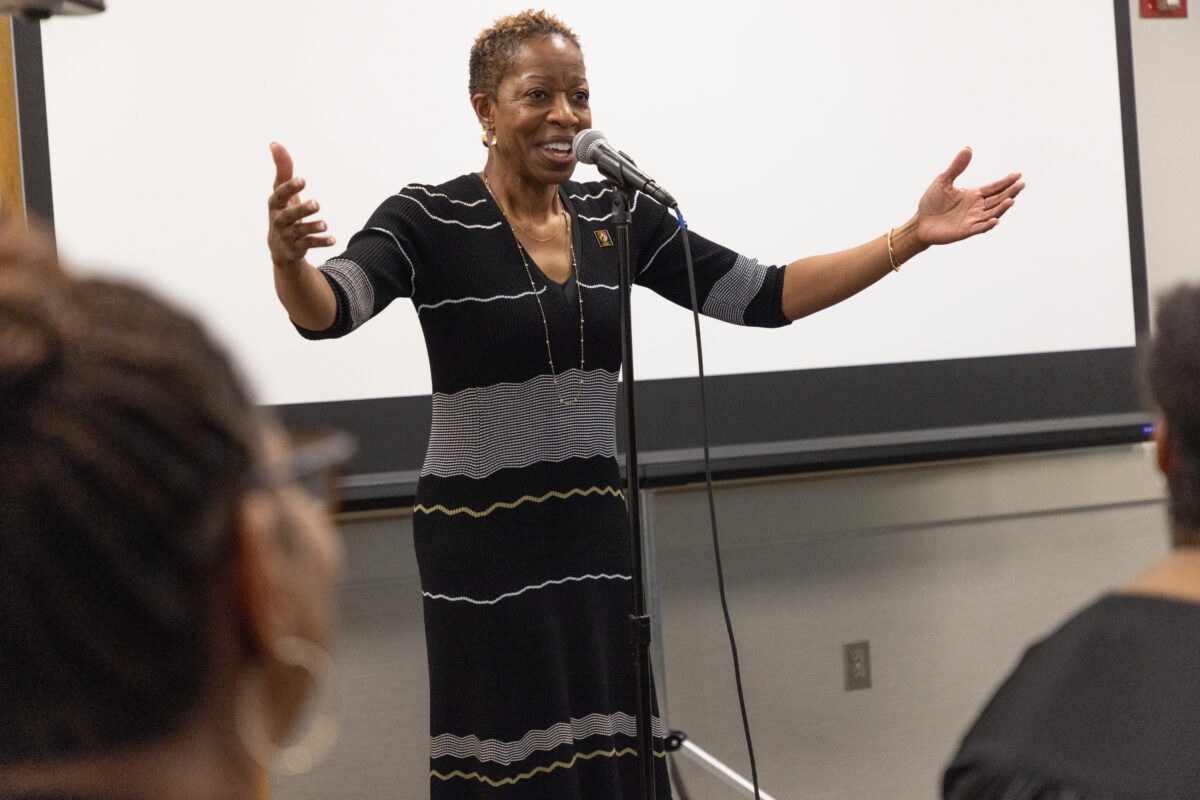
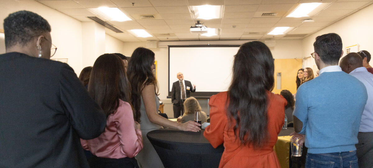
Left: Manfred Van Dulmen, UMBC’s provost, addresses attendees at the reception to celebrate the FIRST faculty. Jim Kaper, professor of microbiology and immunology and the UMSOM lead on the grant; Roger Ward, UMSOM provost; and Valerie Sheares Ashby, UMBC president, look on (from left to right). Right: Valerie Sheares Ashby, UMBC president, addresses the FIRST reception attendees. (Melissa Penley Cormier, M.F.A. ’17)
Li’s research straddles neuroscience and behavior. His model system, African cichlid freshwater fish, is useful for studying reproductive and social behaviors, such as courtship rituals and aggression. Many of the mechanisms involved are applicable across a wide range of species.
Carmen Muñoz-Ballester, another new assistant professor of biological sciences at UMBC, is finding her department colleagues and the FIRST cohort to be helpful resources as she navigates her first year.
“The Department of Biological Sciences at UMBC provides a very supportive environment for building a scientific career, and I am very happy to be part of this thriving community,” Muñoz-Ballester says. “Life as a faculty member can be overwhelming at the beginning, and having the opportunity to share challenges and small wins with people in similar situations makes it much better. I am very glad to have my peers to count on in my daily life, and I am committed to making the most of this opportunity and paying it forward to future trainees and colleagues.”
Muñoz-Ballester’s work focuses on sex differences in the response to traumatic brain injury. Severe or repeated brain injuries (such as seen in football players) are getting more attention today, and men suffer more traumatic brain injuries overall, but she notes that women tend to have worse outcomes than men after sustaining less-severe brain injuries. Muñoz-Ballester will work to unpack the neural mechanisms behind the effects of these injuries in both sexes, potentially leading to more effective treatments.
William R. LaCourse, dean of UMBC’s College of Natural and Mathematical Sciences and UMBC lead on the FIRST grant, addresses attendees at the reception. (Melissa Penley Cormier, M.F.A. ’17)
Tackling the biggest challenges
Diana Elizondo and Gretchen Alicea will round out the FIRST cohort at UMBC. Elizondo is an immunologist who uses mice to study signaling pathways involved in inflammation and metabolic disorders like diabetes and obesity. Alicea focuses on how metabolic changes in the environment immediately surrounding malignant tumors are related to the risk of metastasis. Jose Lemme, Alisha Blazer, and Amed Outtara have been brought on board at UMSOM.
William R. LaCourse, CNMS dean, is thrilled to welcome these new members of the faculty to UMBC. Their research interests include some of the most pressing issues society faces today, like cancer and obesity. These researchers are also approaching study areas that may deserve more attention from innovative angles.
“Thank you for choosing our institutions to be a part of your academic careers. We want you to know that your success goes beyond the UM FIRST program and even beyond tenure. Your success is that you always find joy in research, mentoring, teaching, and life,” LaCourse said at the reception. “This room is full of individuals who are there for you.”
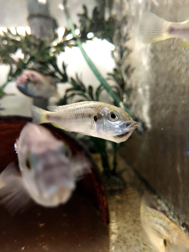
African cichlids, a group of thousands of freshwater fish species, present a wide range of distinctive mating and parental care behaviors. However, the mechanisms that drive these behaviors are poorly understood. Cheng-Yu Li, assistant professor of biological sciences and a member of the UM FIRST faculty cohort, and colleagues have discovered a new clue in this mystery and published their results in Current Biology. A wide range of vertebrate species exhibit versions of these behaviors, so Li and colleagues hope their research will contribute to a broader understanding of the neural mechanisms that are behind them.
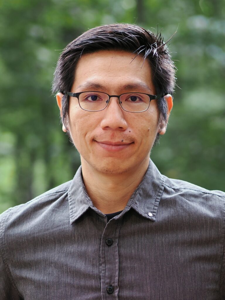
Cheng-Yu Li (courtesy of Li)
Using an African cichlid species, Astatotilapia burtoni, asthe model organism, the authors identified that a group or neurons called ciliated olfactory sensory neurons (OSNs) are the key type of cell responsible for detecting female reproductive pheromones. They further pinpointed a specific pheromone receptor, Or113a, that regulates males’ reproductive behavior.
In male cichlids, sensory neurons expressing the Or113a receptor detect cues derived from a pheromone called prostaglandin F2ɑ, or PGF2ɑ. PGF2ɑ functions both as a female hormone that regulates female mating behavior and as a pheromone that attracts males when females are reproductively receptive. Knocking out their Or113a receptors limited males’ preference for water taken from near a sexually receptive female. Silencing the males’ relevant sensory neurons by knocking out a protein called Cnga2b eliminated the preference entirely.
PGF2ɑ on its own is attractive to males in some fish species, such as zebrafish and goldfish, however, but male A. burtoni cichlids are insensitive to PGF2ɑ. This suggests that PGF2ɑ must be converted into a different chemical in reproductive females or that more than one pheromone is involved in the male attraction response. That makes evolutionary sense, Li explains, because many species of cichlids and other fish living in the African Great Lakes may use PGF2ɑ for pheromone signaling. If it was the only signal, there would be a high probability of interbreeding between cichlid species, or a male could mistakenly approach females of predator species. Using more than one signal would solve this problem. Li and colleagues are actively working on finding additional pheromone clues to further unravel this complex signaling system.
A puzzle worth brooding over
Discovering a neurological mechanism driving males’ attraction to females was exciting for Li, but an unusual observation during their trials added to the study’s impact. Many cichlids “mouth brood,” or carry their eggs in their mouths for several weeks until the larvae can swim on their own. In most species, including the one used in Li’s study, the female is the exclusive mouth brooder. However, Li’s study found that males with mutations in the genes of cnga2b or or113a would take eggs into their mouths as a female would. But only 30 percent of the males who picked up eggs would mouth brood them until hatching; the other 70 percent immediately swallowed the eggs.
Li and colleagues had expected the males with mutated receptors to alter their preference for water containing female pheromones, but the mouth brooding was a surprise. “That was kind of outside of our expectations,” Li says. “It’s something that we had never heard before, and we didn’t even predict that would happen.”
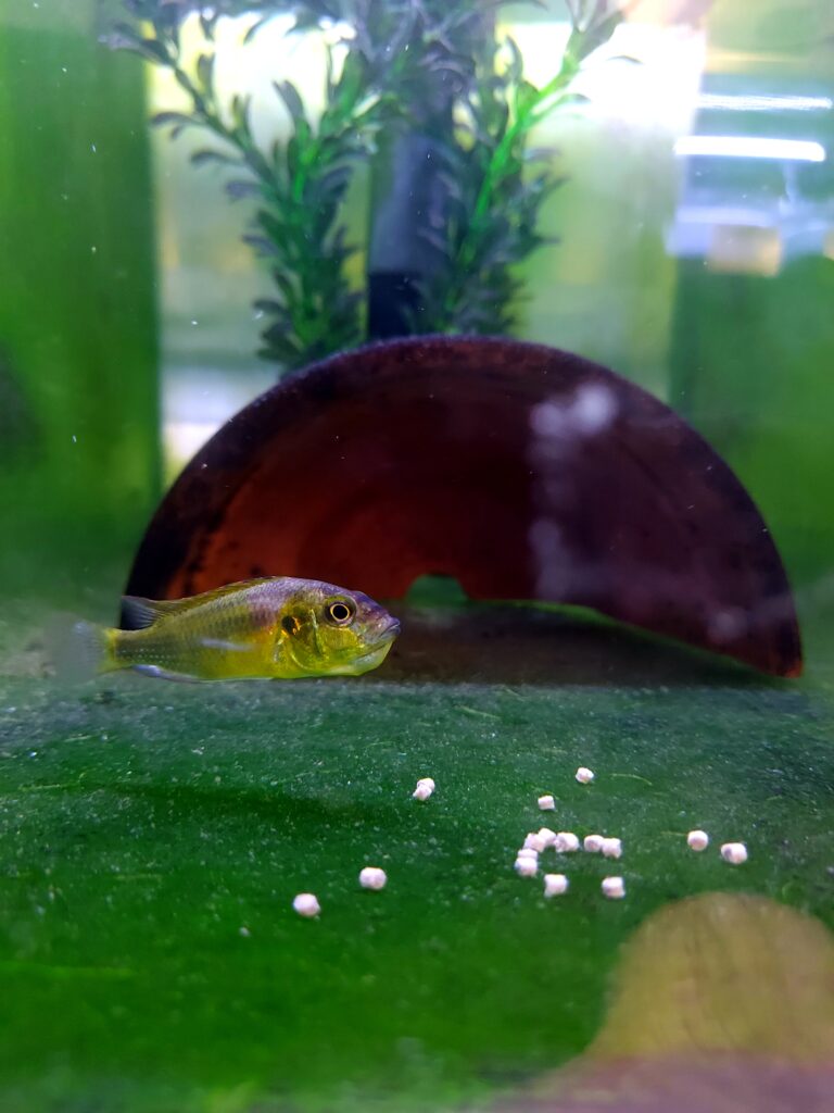
Left: A female A. burtoni holds eggs in her mouth. Right: A male A. burtoni, the cichlid species Li studies, carries eggs in his mouth and ignores nearby food pellets. (Photos courtesy of Li)
To explain the male mouth brooding, “Our first guess is that the PGF2ɑ-derived pheromone cues play dual roles,” Li says. “The first role is to attract males during mating. But it may also inhibit male mouth-brooding behavior. So once you knock out this receptor, this circuit turns on in the males, and they become mouth brooders as well.” In other words, mouth brooding may be the default behavior in both species, but some mechanism driven by pheromones normally turns it off in males.
Supporting this hypothesis, Li and his colleagues found that in blackchin tilapia, a different cichlid species where males normally mouth brood the eggs until hatching, the males may have a naturally occurring mutation in the same Or113a gene. That supports the idea that the phenomenon of male mouth-brooding, and the mechanism that regulates it, “ is a regular mechanism in the field, not just some weird example we saw in the lab,” says Li.
The next question is what additional cues may be required to induce full-term mouth brooding. Li and colleagues are investigating.
Connections across species
Cichlids have a complex social status system, demonstrate significant parental care, and present a diverse set of distinctive mating behaviors, traits they have in common with a wide range of species. Even some of the mechanisms are shared across large evolutionary distances. For example, the hormone prolactin drives parental care in fishes, mice, and even humans.
“We are using this simple model in cichlids, which are easy to breed and easy to study in the lab, and exploring mechanisms that may have broader biological relevance. It’s a wonderful model where we can bridge ecology and evolution concepts with neuroscience,” Li says. “By gaining deeper insights into what regulates parenting behavior, we can identify key neurons and genes in the brain involved in this process, with the goal of uncovering mechanisms that may be shared across various species.”
UMBC at the Universities at Shady Grove finds itself uniquely situated—at home within the country’s third largest biotech hub where the demand for highly skilled workers is growing. These Retrievers are filling specialized roles after graduating with the training they need to succeed in this booming industry.
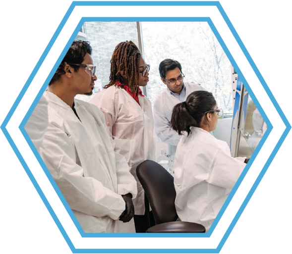
It’s an early Friday morning in the Biological Sciences and Engineering Building and nine students, working in small groups, are bustling back and forth between fume hoods and a large centrifuge. They carefully swish flasks containing cells and growth medium. The smell of sanitizing alcohol pervades the space. Neon orange test tube racks and turquoise tube caps stand out within the sterile white look of the work benches, hoods, and lab coats.
As the cells use oxygen to release energy, they produce carbon dioxide. The CO2 turns the solution yellow, indicating successful cell growth. After the experiment, students sterilize their solution with bleach for disposal; in response to the alkalinity, it blooms to a brilliant magenta. In another room, one student offers tips to her classmate on how to use a pipet more effectively, and the instructor flits between groups, answering questions as needed—but for the most part, the students operate confidently on their own.
This is a regular day in Biotechnology 303: Applied Cell Biology in UMBC’s Translational Life Science Technology (TLST) program. The program, offered by UMBC’s College of Natural and Mathematical Sciences (CNMS) exclusively at the Universities at Shady Grove (USG), a multi-institution education facility in Rockville, Maryland, is designed to offer hands-on training to prepare students for careers in the booming biotech industry in USG’s backyard.
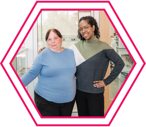
Elizabeth Friar (left) and Samantha Petros ’23 have stayed in touch since Petros graduated.
A clear direction
It was the promise of experiential work with real-world applications that drew Samantha Petros ’23 to the TLST program. “I really liked the convenience of the USG campus, and a lot of the courses were really focused on hands-on learning,” she says. Without really knowing what the work would look like before she started the program, “I was able to try out a lot of different things in a very low-stakes environment and get an idea as to what I found interesting.”
That turned out to be cell culture work. Applied Cell Biology with Elizabeth Friar, lecturer and undergraduate program director in TLST, “cemented that benchwork is what I enjoy doing,” Petros says. From then on, “There was a clearly defined arrowhead in the direction I wanted to go.”
That was a big shift from where she started, as a film major at Montgomery College (MC). A virology course to meet her science requirement turned her on to MC’s biotech associate degree, and then she transitioned to the TLST program at UMBC. After learning some basic lab techniques in TLST classes, she landed a part-time job, where she carried out entry-level manufacturing tasks for malaria vaccines. Today, Petros is thriving as a cell culture specialist studying malaria at Axle Informatics, a contractor for the National Institute of Allergy and Infectious Diseases.
The position she’s in now traditionally requires a master’s degree, which she doesn’t have—yet, anyway. “Knowing that I have valuable skill sets from TLST—working in manufacturing, getting good connections with my professors, and going to the networking events that TLST offers—all of that compounds and has got me to where I am now,” Petros says. “I genuinely believe that I wouldn’t be here if it weren’t for all of those factors, with TLST obviously being the biggest one.”
Location, location, location
TLST is a young program, but it is growing quickly. The first class—just two students—arrived in fall 2019. Friar joined in summer 2020 and began laying the groundwork for the program’s eventual growth. Today, “It’s humming along,” she says. There are currently 50 students who have declared TLST as their major. “Now we’re thinking about what we can do next. How can we expand?”
TLST is unique as the only undergraduate STEM program at UMBC without a footprint on the main campus. The location choice at USG is intentional. The Capital Biohealth Region, encompassing the District of Columbia, Virginia, and Maryland, is the third most competitive biotech hub in the country—and Montgomery County, Maryland, is the hub of the hub, boasting more than 350 life science companies and located a stone’s throw from the NIH, FDA, and National Institute of Standards and Technology (NIST). For TLST alumni, the employment options are seemingly limitless: 80 percent of American pharmaceutical companies are within a two-hour drive, and they can expect a nearly 23 percent growth rate in the biotech industry over the next 10 years—well above the national average.
As Friar puts it, “Regenxbio is down the street. MacroGenics is down the street. MilliporeSigma is opening their new lab right next door, and they’ve now hired eight of our alumni. Soon we’ll have an NIH vaccine lab right down the street. ATCC is right down the street.” All these biotech companies need qualified workers—and TLST is providing them.
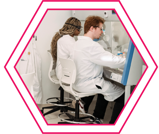
Steven Schaffer (right) and Princess Nyamali work under a biosafety cabinet in Elizabeth Friar’s Applied Cell Biology course.
Training lifetime learners
Jeff Galvin, CEO of American Gene Technologies (AGT), has hired TLST alumni and would hire more. “The TLST program seems to provide a great overview of not just biotechnology techniques,” he says, but it gives students “an understanding of the business in general. I find that UMBC students lean toward being self-motivated problem-solvers. It seems that the administration from the top all the way down promotes that idea of creating value through creative, hard work.”
Titina Sirak ’20, TLST, had a major impact at AGT. She established a brand-new laboratory certified to handle human cells from scratch, Galvin says, “And that’s not easy—there’s a lot of regulations associated with that, even economics.” While she wasn’t an expert at first, “she was able to do enough things right that the project came to life.”
Galvin emphasizes the importance of “learning how to learn,” saying, “Things are changing so fast, that it’s the folks who can adapt and become lifetime learners who are going to be the most successful.And that’s something I saw from the students coming out of TLST.”
A vision fulfilled
So far, TLST has graduated 25 students, and 94 percent of them were employed in the biotech industry within three months of walking across the Commencement stage. “We’ve had a number of really standout students who have gone on to do really great things, so I’m very excited about how it’s going,” Friar says.
Merryll Kallungal ’24 works under a biosafety cabinet at ATCC. Interns Jason Bose (left) and Tamilore Akinde (second from left) and an ATCC lead scientist observe.
So is William R. LaCourse, CNMS dean, which offers the TLST program. He envisioned the program years before the first two students walked onto the USG campus, and it came together with input from faculty at Montgomery College and five departments at UMBC.
“TLST is an innovative and practical education that combines ‘know-what’ and ‘know-how,’” says LaCourse. “It is a highly flexible program with various pathways to serve the ever-changing needs of a growing industry. TLST is where the silos of disciplines break down and cross-disciplinary knowledge is the goal—both in content and practice.”
The program is shaping up just as LaCourse and Annica Wayman ’99, mechanical engineering, and former associate dean for Shady Grove Affairs in CNMS who led TLST’s launch, had hoped. The state of Maryland is growing its biotech hub while UMBC students are gaining the skills they need to succeed in the workplace and getting well-paying jobs, and the student population is growing.
Connections that build confidence
Just as important as high-demand lab skills, students and alumni value the relationships they’ve formed with faculty. “Every teacher I had was very supportive, very understanding, very willing to work with me,” Petros says. “I think it’s important to get your money’s worth out of college, and I definitely feel like I got that and more in TLST because of the support network that I had there and my teachers.”
Petros is still in touch periodically with Wayman and Friar. “Dr. Wayman was a shining inspiration. She’s just a wonderful person. She’s so knowledgeable and always willing to help,” Petros says. The support she has and the success she’s found now have inspired Petros to look back and help those coming up behind her. In addition to USG resources, as a student Petros attended networking events offered by BioBuzz, a community resource for biotech industry professionals and job seekers in the Capital Biohealth Region. In fact, it’s how she found her current role.
Today Petros is a BioBuzz Ambassador. “I wish I had known about BioBuzz when I first started in biotech,” she says. “Now I want to be that advocate for younger students or people who are just starting in science.”
A strong foundation
“Our students come to us because they want to make a difference in people’s lives, but they don’t necessarily want to go to medical school,” Friar says. It doesn’t hurt that “we have some really spectacular facilities that are a real draw for students,” she adds.
The Biological Sciences and Engineering Building opened at USG in fall 2019 and boasts beautiful modern laboratories and classrooms plus plenty of comfy nooks for meeting with a study group or just relaxing. TLST has also received over $1 million dollars from the National Institute for Innovation in Manufacturing Pharmaceuticals, some of which has supported further upgrades and additions to the teaching laboratories.
A flow cytometer, a machine that can detect properties of interest in up to 10,000 cells per minute, will enable a new mixed undergraduate/graduate course in flow cytometry launching next spring, for example. And equipment to practice skills like protein purification, biomanufacturing processes, and cell culture create rare opportunities for undergraduates. A required TLST course covers the wide range of instrumentation you might find in a modern biotech laboratory.
It’s normal for students to start off nervous when they use complicated and expensive equipment or work with human cells for the first time. “But by the end, they are old hands, and I think they would feel comfortable tackling anything in that lab on their own,” Friar says. “And it’s because the curriculum is really well scaffolded. We add skills as they go along, and then we repeat the old skills. The program is set up to foster growth and independence.”
Princess Nyamali stepped away from the centrifuge for a moment during Biotechnology 303 to share that the TLST coursework “is a really good foundation.” She’s currently completing an internship at NIST, and “everything I’m learning in this cell culture class is stuff I’m doing at NIST.” While it might have been daunting at first, trying so many different things across the TLST curriculum “helps you know that you want to do it,” she says, in addition to showing potential employers that you can.
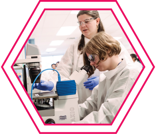
TLST student Fae Switzer works at the microscope with guidance from her mentor, Emma Todd, at ATCC.
Thinking ahead
Although the two-year program initially targeted transfers from regional community colleges, “we were getting so much interest from students on the Catonsville campus, we went ahead and put it on the list of majors for freshmen applying to UMBC,” Friar says. “That’s been the fastest growing cohort in our major.”
Steven Schaffer transferred to TLST after starting on the main campus in bioinformatics. Now he is in the bioinformatics track within TLST; the other option is a biomanufacturing track. Schaffer likes that there is a strong cohort connection because everyone takes mostly the same classes together. “I would recommend TLST. It’s a growing field and the skills you learn are very versatile,” Schaffer adds.
Schaffer hopes to eventually take on a role that leans into engineering at a place like Northrop Grumman. But before that, he’s eyeing UMBC’s Master of Professional Studies in biotechnology. The M.P.S. degree offers advanced instruction in the life sciences, plus coursework in regulatory affairs, leadership, management, and financial management. Friar describes it as “a cross between a science master’s and an M.B.A.”
Academic decathletes
Soon, the confident students in Friar’s cell biology class will arrive in labs across Montgomery County, the capital region, and beyond. They will contribute to drug discovery and production, design manufacturing processes, and, eventually, lead teams and make strategic decisions for major biotech companies.
Jeff Galvin, the ATG CEO, referred to TLST alumni as “academic decathletes.” Like decathletes, they have an array of skills and perform all of them admirably. But rather than medals, they’re seeking an opportunity to contribute to positive change through their work. They’ll translate basic science into diagnostic tests, treatments for disease, and more to improve the lives of their neighbors in Maryland and those in need around the world.
ATCC’s SPARC internship program offers opportunities for TLST students to conduct meaningful work in the biotech industry. Merryll Kallungal (far left) transitioned from her internship to a full-time role at ATCC this summer.
SPARC-ing Rewarding Careers
Just 10 minutes from USG in Gaithersburg, Maryland, biotech company ATCC has capitalized on the TLST talent pipeline. Their Student Partnership and Research Collaboration (SPARC) Program requires USG enrollment and offers qualified students paid, part-time career opportunities throughout the academic year at ATCC’s research facility. As the leading developer and supplier of authenticated cell lines, microorganisms, and associated data for academia, industry, and government, a successful internship at ATCC is a feather in the cap for students pursuing a TLST degree.
Of the seven interns selected for the SPARC program this year, four are TLST students—and they are thriving. The interns praised their SPARC experience while donning lab coats, shoe booties, safety glasses, and gloves in one of ATCC’s labs—where keeping employees safe and protecting lab samples and products from contamination is mission critical.
“I love the TLST program!” exclaimed Fae Switzer, who has wanted to pursue a career in biotech since she learned about CRISPR, the gene-editing platform, as a child. Switzer, a SPARC associate biologist in the microphysiological unit, notes that things she learned in her UMBC classes prepared her for the internship, and things she’s learning at ATCC now are helping her in her classes.
Jason Bose, a SPARC associate biologist on the microbiology team, knew he was interested in biology, but not medical school. When Bose learned about TLST, he thought, “Wow, this aligns with what I want to do,” he says. “It’s been really great so far.” He even uses the basic coding skills and tricks for Excel spreadsheets that he learned in class—which at the time he wasn’t so sure about. “Yep, I use them all the time at ATCC,” he says.
Tamilore Akinde completed an internship last summer at ATCC’s headquarters in Manassas, Virginia, and now she is a SPARC intern. “I like that TLST teaches multidisciplinary skills,” she says. Being able to pivot toward different opportunities that come her way is valuable, she adds.
“The hands-on lab training provided by the TLST program has prepared students for impactful careers at ATCC,” shares Ruth Cheng, general manager and senior vice president for research and industrial solutions at ATCC. “Witnessing students like Merryll Kallungal transition from the SPARC program to a full-time role in cryobiology R&D is a testament to the power of this collaboration.”
After interning at ATCC, Kallungal graduated from TLST in May and assumed a full-time associate biologist role in the cryobiology group in June. The lab techniques she learned in TLST came in handy at ATCC, and she has already learned a lot of new things, too. “I absolutely love what I do,” she says.